Background on Disease
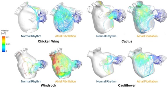
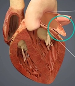
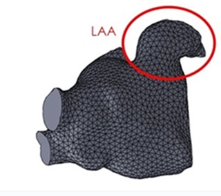
About 9 million people in the United States are diagnosed with atrial fibrillation (AF), a common cardiac condition characterized by disorganized electrical signals that cause rapid and irregular heart muscle contractions. This abnormal rhythm disrupts the efficient flow of blood through the heart, particularly affecting a 2-4 cm finger-like sac protruding from the left atrium called the left atrial appendage (LAA). The LAA acts as a small reservoir for blood, contributing to the overall mechanical function of the left atrium by helping to regulate heart volume and pressure.
 However, during AF, the erratic heart rhythm leads to sluggish blood flow within the LAA, creating an environment conducive to thrombus (blood clot) formation. The LAA's complex shape and slower blood flow make it a prime site for clots to develop. These clots pose a significant risk because they can dislodge and travel through the bloodstream to the brain, causing a stroke. In fact, over 90% of stroke-causing blood clot events in individuals with AF originate from the LAA.
Due to this high risk, managing AF often involves strategies to prevent clot formation in the LAA. This includes the use of anticoagulant medications, which thins the blood to reduce the likelihood of clot formation. Additionally, procedural interventions such as LAA closure devices (e.g., the WATCHMAN device) or surgical occlusion are employed to seal off or remove the LAA, thereby eliminating the primary site for clot formation.